Global Reach
번역샘플 - 한영 번역
품질 및 시기 적절한 투고물을 제공하는 것은 급선무로 특히 조절 의뢰의 경우 더욱 필요하다.
이나고는 주제별 전문가 리뷰어, 번역가, 저자, 교정자로 이루어진 특별한 팀과 함께 더불어 귀하의 언어로 24 시간 전문 프로젝트 관리자를 제공함으로써 정시에 고품질의 투고물을 제공합니다. 저희는 단어 대 단어 번역을 뛰어 넘어 최첨단 기술과 엄격한 프로세스를 통해 귀하의 주제 영역의 뉘앙스까지 살린 고품질 번역을 제공합니다.
아래는 저희의 우수한 번역본의 샘플들입니다.
생명과학, 생물자원학, 농업 관련 학과
인간 배아줄기세포 (human embryonic stem cell; hESC)는 착상 전 배반포 의 내부세포덩어리 (inner cell mass; ICM)로부터 확립된 세포로서, 적절한 배 양조건의 환경에서 외배엽, 중배엽, 내배엽의 삼배엽 (three germ layer)의 다 양한 세포로 분화 가능한 전분화능 (pluripotency)세포이다 (1, 2). 인간 배아줄 기세포가 확립 된 이래로 이와 같은 배아줄기세포의 특성을 이용하여 인간의 초기발생 및 분화에 관여하는 각종 유전자 및 분화 유도인자들의 작용기전 등 을 밝히고자 하는 다양한 연구가 이루어지고 있다. 또한 질병을 치료하는 세 포치료제로 활용하기 위하여 인간 배아줄기세포를 심근세포 (3, 4), 신경 및 신경교세포 (5, 6), 조혈모 전구체 (7, 8) 등의 특정한 세포로 분화를 유도하는 연구가 활발히 이루어지고 있다.
일반적으로 배아줄기세포의 분화는 원시 내배엽 유사세포 (primitive endoderm like-cell; PrE like-cell)가 다수 포함되어있는 자연분화 된 다양한 형태 (9)와 특 성 (2, 10, 11)을 지닌 자연분화 (spontaneous differentiation)가 있으며, 어떠한 성장인자나 화학물질을 처리하지 않고 단지 배양 조건에 따른 물리적인 변화 에 의해 자발적으로 분화되는 배상체 형성 (embryoid body formation)이 있 다 (11). 또한 다양한 세포분화에 관련된 성장 인자들을 처리하거나 특정한 화학물질을 처리하여 원하고자 하는 세포로 배아줄기세포의 분화를 유도하 는 유도분화 (guided differentiation)가 있다 (12).
분화를 위해 사용된 가장 일반적인 방법은 지지세포층으로부터 인간 배아 줄 기세포를 분리하여 부유 상태에서 배양을 하는 것으로, 부유배양 (suspension culture) 후에 배상체 (embryoid body; EB)라 불리는 3차원 구조의 구형을 형 성하게 되며, 3차원 구조 형성 동안에 분화가 시작된다. 배상체는 서로 상호작 용을 하고 분화를 유도하며, 삼배엽성 세포로 구성되어 여러 계통으로 종합적 인 분화가 이루어진다. 그리고 배양 초기에 다층의 세포 덩어리로 이루어진 단순 배상체 (simple EB)가 형성되며, 배양 기간이 길어짐에 따라 세포 덩어 리 가운데에 강 (cavity)이 형성되고 (cavity EB), 그 이후에는 용액이 차있는 포낭 배상체 (cystic EB)가 형성된다 (11, 13). 이와 같이 배양시기에 따라 형 태와 특성을 구분한다는 보고가 있다. 그러나 실제로 배상체를 형성시킬 때 항상 일관성 있는 형태와 특성을 가진 배상체가 출연하는 것은 아니며 (14,15), 이 현상에 대한 기전은 아직 알려져 있지 않다.
A native translator who is also a subject expert translates the original document
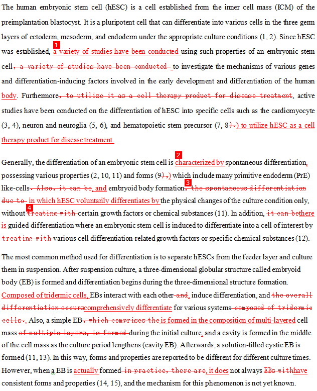
The human embryonic stem cell (hESC) is a cell established from the inner cell mass (ICM) of the preimplantation blastocyst. It is a pluripotent cell that can differentiate into various cells in the three germ layers of ectoderm, mesoderm, and endoderm under the appropriate culture conditions (1, 2). Since hESC was established, a variety of studies have been conducted using such properties of an embryonic stem cell to investigate the mechanisms of various genes and differentiation-inducing factors involved in the early development and differentiation of the human body. Furthermore, active studies have been conducted on the differentiation of hESC into specific cells such as the cardiomyocyte (3, 4), neuron and neuroglia (5, 6), and hematopoietic stem precursor (7, 8) to utilize hESC as a cell therapy product for disease treatment.
Generally, the differentiation of an embryonic stem cell is characterized by spontaneous differentiation, possessing various properties (2, 10, 11) and forms (9) which include many primitive endoderm (PrE) like-cells, and embryoid body formation in which hESC voluntarily differentiates by the physical changes of the culture condition only, without certain growth factors or chemical substances (11). In addition, there is guided differentiation where an embryonic stem cell is induced to differentiate into a cell of interest by various cell differentiation-related growth factors or specific chemical substances (12).
The most common method used for differentiation is to separate hESCs from the feeder layer and culture them in suspension. After suspension culture, a three-dimensional globular structure called embryoid body (EB) is formed and differentiation begins during the three-dimensional structure formation. Composed of tridermic cells, EBs interact with each other, induce differentiation, and comprehensively differentiate for various systems. Also, a simple EB is formed in the composition of multi-layered cell mass during the initial culture, and a cavity is formed in the middle of the cell mass as the culture period lengthens (cavity EB). Afterwards, a solution-filled cystic EB is formed (11, 13). In this way, forms and properties are reported to be different for different culture times. However, when a EB is actually formed, it does not always have consistent forms and properties (14, 15), and the mechanism for this phenomenon is not yet known.

Comments
감수자– 명료성:문맥 상의 명료함을 위해 좀더 자연스럽게 수정 감수자– 명료성:문장의 명료성을 위해 추가 감수자–누락:누락 된 부분이 추가됨. 원문 "자발적으로 분화되는" 감수자– 유창성:“treating with” (처리하다)는 문장의 명료성을 높이고 장황성을 줄이기 위해 삭제

원어민 에디터-가독성: 글의 간결성을 위해 수정 감수자–명료성:문맥 상의 명료함을 위해 좀더 자연스럽게 수정 감수자– 명료성:문장의 명료성을 위해 추가 원어민 에디터-약어: 약어가 처음 쓰일 때에는 그 뜻을 풀어써야 함 감수자–누락:누락 된 부분이 추가됨. 원문 "자발적으로 분화되는" 감수자–유창성:“treating with” (처리하다)는 문장의 명료성을 높이고 장황성을 줄이기 위해 삭제 원어민 에디터-관사:단어의 첫 음이 모음이라면 관사 ‘an’을 사용해야하나 ‘a’가 사용됨 원어민 에디터-개선:원문의 표현을 개선하기 위해 수정이 이루어짐
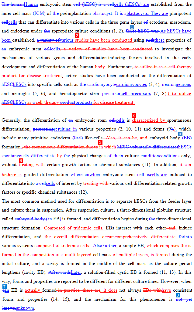
Human embryonic stem cells (hESCs) are established from the inner cell mass of preimplantation blastocysts. They are pluripotent cells that can differentiate into various cells in the three germ layers ectoderm, mesoderm, and endoderm under appropriate culture conditions (1, 2). As hESCs have been established, various studies have been conducted using these properties of embryonic stem cells to investigate the mechanisms of various genes and differentiation-inducing factors involved in the early development and differentiation of the human body. Furthermore, active studies have been conducted on the differentiation of hESCs into specific cells such as cardiomyocytes (3, 4), neurons and neuroglia (5, 6), and hematopoietic stem cell precursors (7, 8) to utilize hESCs as cell therapy products for disease treatment.
Generally, the differentiation of embryonic stem cells is characterized by spontaneous differentiation, resulting in various properties (2, 10, 11) and forms (9), which include many primitive endoderm like-cells, and embryoid body (EB) formation, in which hESCs spontaneously differentiate by physical changes in culture conditions only, without certain growth factors or chemical substances (11). In addition, there is guided differentiation when embryonic stem cells are induced to differentiate into cells of interest by various cell differentiation-related growth factors or specific chemical substances (12). The most common method used for differentiation is to separate hESCs from the feeder layer and culture them in suspension. After suspension culture, a three-dimensional globular structure called an EB is formed, and differentiation begins during three-dimensional structure formation. Composed of tridermic cells, EBs interact with each other, induce differentiation, and comprehensively differentiate into various systems. Further, a simple EB is formed in the composition of a multi-layered cell mass during the initial culture, and a cavity is formed in the middle of the cell mass as the culture period lengthens (cavity EB). Later, a solution-filled cystic EB is formed (11, 13). In this way, forms and properties are reported to be different for different culture times. However, when an EB is actually formed, it does not always have consistent forms and properties (14, 15), and the mechanism for this phenomenon is still unknown.


 학술출판번역
 论文翻译
论文翻译 学術翻訳
学術翻訳 English
 學術翻譯
學術翻譯 traduzione cartacea
 Papierübersetzung
 traduction papier
 papieren vertaling
 ترجمه مقاله
 แปลกระดาษ
 бумажный перевод
 ترجمة ورقية
 makale çeviri
 Tradutor de Artigos


